
Chronic Obstructive Pulmonary Disorder (COPD)
Definition
- Heterogenous lung condition characterized by chronic respiratory symptoms (dyspnea, cough, sputum production &/or exacerbations)
- Due to abnormalities of the airways (bronchitis/bronchiolitis) &/or alveoli (emphysema)
- Causing persistent, often progressive airflow obstruction.
Pathophysiology
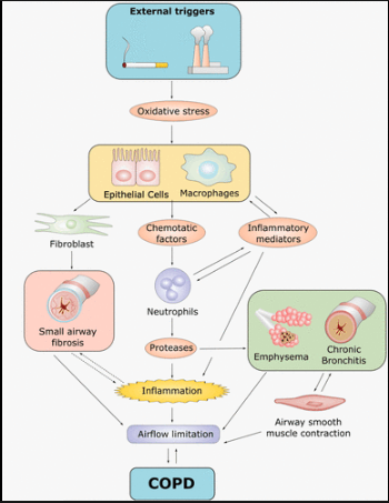
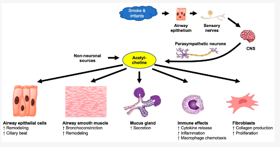
As seen above, acetylcholine plays a role in the pathophysiology in COPD and thus often time in the management of COPD we see the usage of SAMA or LAMA but not so in asthma management.
- Repeated exposure to cigarette smoke & other noxious particles -> chronic inflammation & structural changes in the airways, lung parenchyma & pulmonary vasculature.
- The inflammatory response—predominantly involving neutrophils, macrophages and CD8⁺ lymphocytes—differs from that in asthma.
- Chronic inflammation –> Mucus hypersecretion + dynamic small airway collapse + emphysema –> expiratory airflow limitation (inability of the airway to remain open during expiration) –> air trapping, lung hyperinflation, gas exchange abnormalities & progressive pulmonary HTN –> cor pulmonale
- Respiratory infections and environmental pollutants may amplify inflammation and trigger exacerbations.
Risk factors
COPD develops through GETomics: the interaction between genetic background (G) and environmental risk factors (E) over the lifetime (T).
These risk factors include :
- Genes (severe alpha-1 antitrypsin enzyme deficiency –> panlobular emphysema)
- Exposure to particles
– Tobacco smoke (dose-related)
– Organic & inorganic occupational dusts
– Indoor air pollution from heating & cooking with biomass in poorly ventilated dwellings.
– Outdoor air pollution (air pollution from motor vehicle emissions is a/w decrease in lung function) - Lung growth & development : Early-life factors may result in a lower maximally attained lung function
- Asthma & airway hyper-reactivity
- Chronic bronchitis
- Oxidative stress
- Gender : Female may be more susceptible to the effects of tobacco smoke
- Respiratory infections (vicious cycle hypothesis)
– A h/o severe childhood respiratory infection is associated with reduced lung function & increased respiratory symptoms in adulthood.
– e.g. chronic bronchial infection (especially with Pseudomonas aeruginosa), TB, HIV - Socioeconomic status (inversely related)
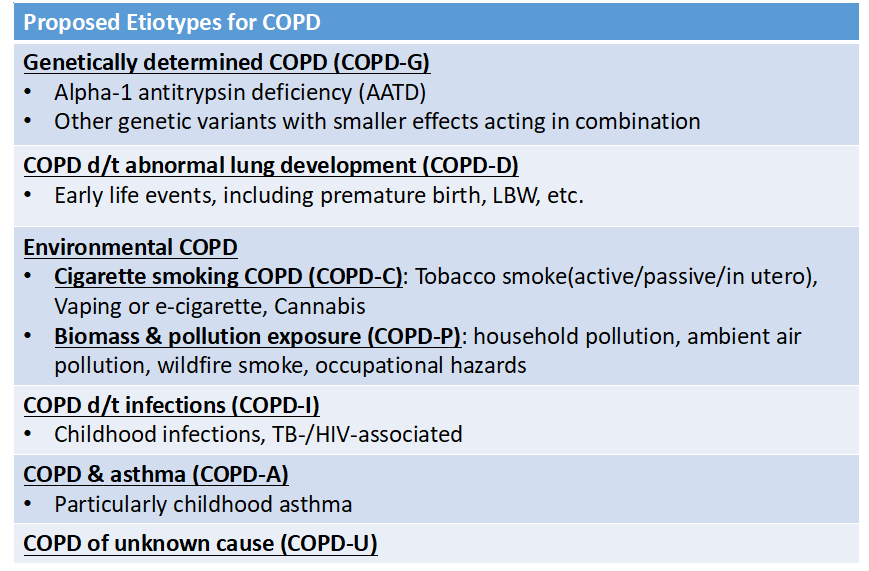
The risk factors can be categorized into few proposed etiotypes for COPD in the GOLD 2026 guidelines
Approach
1. History
a) Symptoms – may fluctuate from day to day
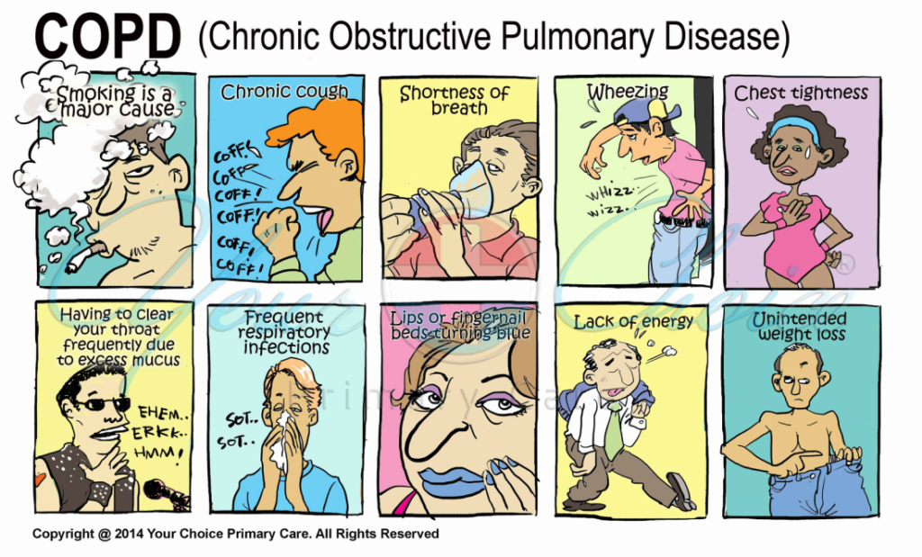
- Dyspnea (hallmark) – progressive, persistent & worse with exercise. It may occur at any stage. Highly prevalent across all stages of airflow obstruction.
- Cough (often the 1st symptoms) – may initially be intermittent, later present daily, with or without sputum
– In severe airflow obstruction, prolonged coughing may rapidly increase intrathoracic pressure and cause cough syncope.
– Coughing spells may also cause rib fractures, which are sometimes asymptomatic. - Sputum production – may be intermittent with periods of flare-up & remission.
- Wheezing & chest tightness – may be present, but their absence does not exclude COPD, nor does their presence confirm asthma.
- Fatigue, Depression &/or anxiety
- Advanced/Severe cases: Weight loss, muscle mass loss, anorexia or ankle swelling (due to cor pulmonale)
b) Severity assessment (discussed in more detailed at subsequent section)
c) Risk factors
- Smoking history – Quantification of tobacco consumption: total pack-years = (no. of cigarettes smoked per day/20) x no. of years of smoking.
- Occupational, household & environmental exposures to other lung irritants
d) Relevant medical history, including :
- Early life events – e.g prematurity, low birthweight, maternal smoking during pregnancy, passive smoke exposure during infancy)
- Asthma, allergy, sinusitis, or nasal polyps
- Respiratory infections in childhood; HIV; TB
- History of exacerbations or previous hospitalizations for respiratory disorder
e) Other relevant history
- Family h/o any respiratory disorder
- Co-morbidities: CVS disease, PSY illness, malignancy, osteoporosis, MSK disorder etc.
- All current medical therapy & its appropriateness
- Impact of disease on psychosocial well-being.
- Social & family support
Clinical indicators (GOLD 2026 guidelines)
- Dyspnea: progressive over time, worse with exercise, persistent
- Recurrent wheeze
- Chronic cough: may be intermittent & may be unproductive
- Recurrent LRTIs
- Presence of risk factors (tobacco smoke, occupational dust, genetic factors etc.)
2. Physical examination
Not usually diagnostic.
Signs of airflow limitation & air trapping (e.g. barrel chest, loss of cardiac & liver dullness, prolonged expiration, ⬇ breath sounds) are usually seen at advanced stage.
Vital signs, especially oxygen saturation (SpO2) should be assessed to detect resting hypoxemia.
- Consider ABG or referral if SpO2 < 92%
Look for complications e.g. malnourishment, cor pulmonale
3. Diagnosis
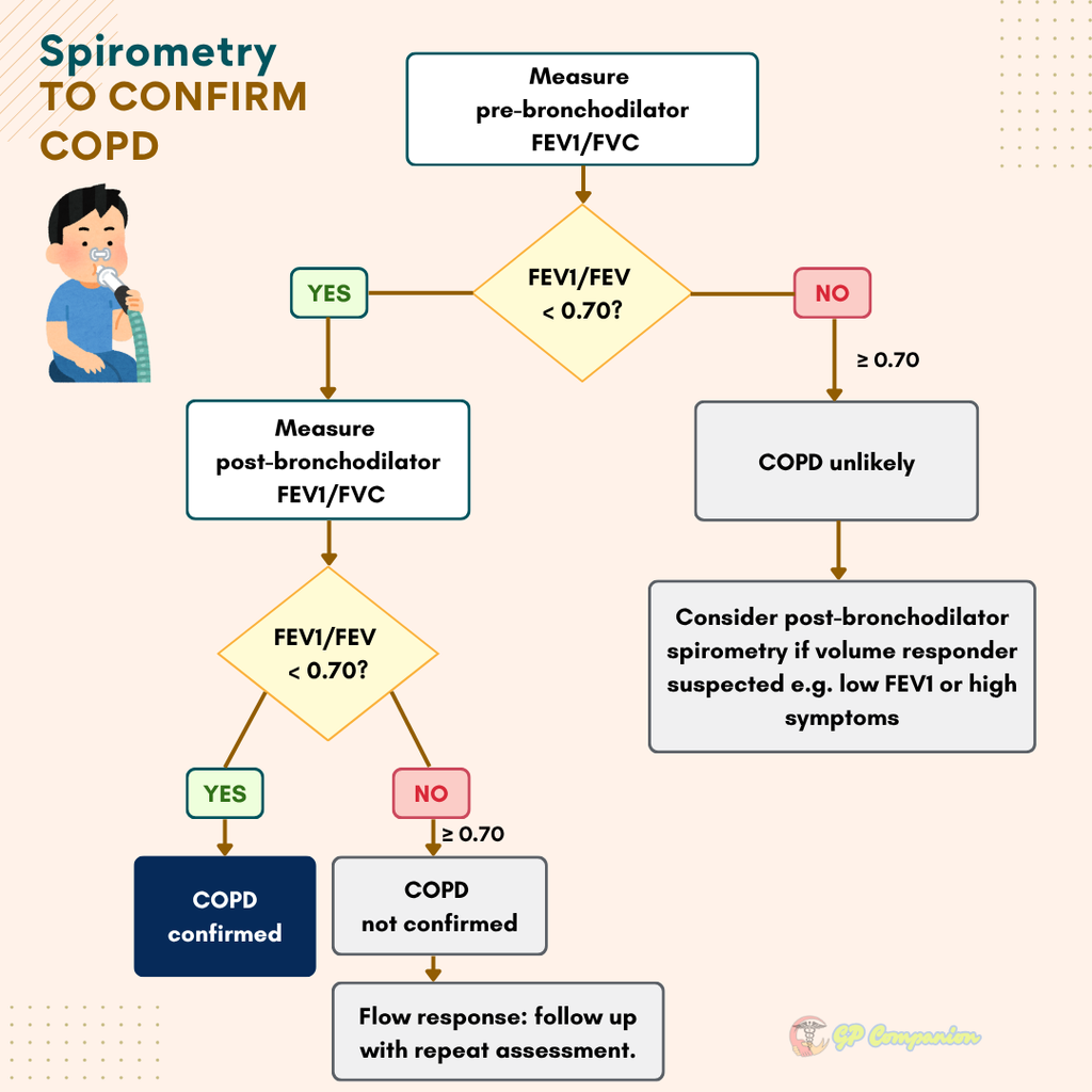
Diagnosis should be confirmed by spirometry showing a post-bronchodilator FEV1/FVC ratio of < 0.7.
- Spirometry should be performed after an adequate dose of inhaled bronchodilator (e.g. 400 ug of salbutamol) in order to minimize variability.
- Peak flow measurements can detect airflow limitation but has poor specificity & correlates poorly with FEV1.
GOLD 2026 recommends using both pre- and post-bronchodilator spirometry approach to confirm the diagnosis of COPD.
Pre-bronchodilator spirometry can be used as 1st step to determine whether symptomatic individuals have airflow obstruction.
Practical approach :
1. Perform pre-bronchodilator spirometry as an initial test.
2. If airflow obstruction is present, confirm COPD with post-bronchodilator spirometry.
- If the post-bronchodilator FEV₁/FVC is 0.60–0.80, repeat spirometry on a separate occasion because of possible biological variation.
- A post-bronchodilator FEV₁/FVC ratio <0.60 is unlikely to rise spontaneously above 0.70.
3. If no obstruction is present on pre-bronchodilator test, post-bronchodilator testing is generally unnecessary unless clinical suspicion remains high, in which case, an FVC volume response may reveal obstruction.
Important consideration
Persistent airflow obstruction is not specific to COPD & may occur in asthma & other conditions. Spirometry findings should be interpreted alongside the clinical features & risk factors.
Assessment
Once diagnosis of COPD is confirmed, subsequent assessment must focus on these fundamental aspects to guide therapy:
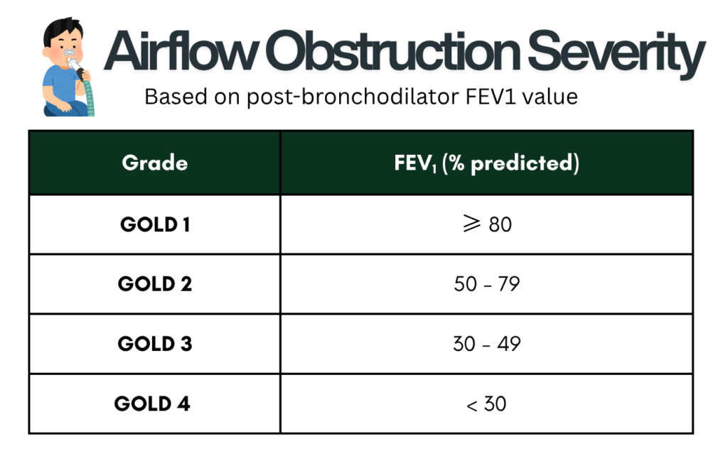
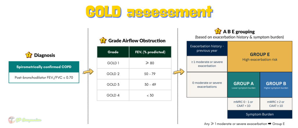
a. Severity of airflow obstruction – based on post-bronchodilator value of FEV1 (GOLD 1 – 4)
b. Symptoms burden
- Symptoms correlate poorly with the degree of airflow obstruction. Thus, validated questionnaire should be used to assess symptom burden.
- Recommended questionnaires :
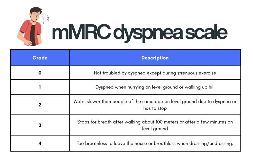
– mMRC dyspnea scale (dyspnea questionnaire) – relates well to other multidimensional health status measures & predicts mortality risk.
– The CAAT/CAT questionnaire (multidimensional questionnaires)
– BODE Index (used to predict mortality which helps with prognosis counseling, not for guiding treatment)
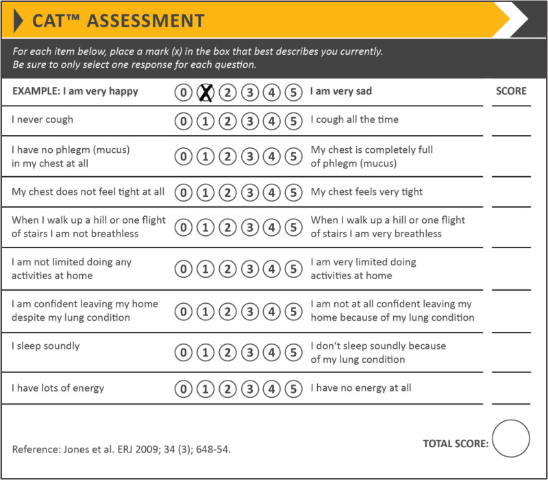
Considering regular treatment, when :
- CAAT ≥ 10 (equivalent to SGRQ score ≥ 25 which is the most comprehensive tool, but more complex to be used in routine practice)
- mMRC ≥ 2 (because mMRC assesses only dyspnea, evaluate for other COPD symptoms even with mMRC < 1)
COPD severity classification based on post-bronchodilator FEV1 & symptoms (Malaysia CPG)
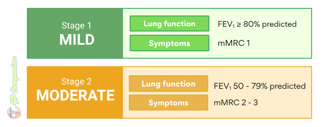
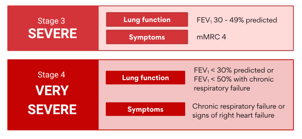
c. Exacerbation risk
- The strongest predictor of future exacerbation is a previous exacerbation, especially :
– > 1 exacerbation requiring steroids &/or antibiotics OR
– Any exacerbation requiring hospitalization. - Even 1 moderate or severe exacerbation requiring corticosteroids, antibiotics or hospitalization may accelerate lung-function decline & worsen prognosis.
d. Blood eosinophil counts
- Recommended by GOLD to guide the use of ICS as part of pharmacological management, as it helps to predict the magnitude of the effect of ICS (in addition maintenance bronchodilator treatment) in preventing exacerbations.
- Higher counts generally reflect greater type 2 airway inflammation, although blood & airway T2 biomarkers do not always correspond closely.
e. Comorbidities
- Comorbidities independently influence hospitalization & mortality, and should be treated according to the relevant guidelines.
- Common multimorbid diseases may include : CVD, metabolic syndrome, osteoporosis, depression & anxiety.
- Significant extrapulmonary (systemic) effects of COPD include weight loss, nutritional abnormalities, & skeletal muscle dysfunction –> can contribute to exercise intolerance & poor health status.
GOLD guideline approach to COPD assessment
Investigations to consider at primary care
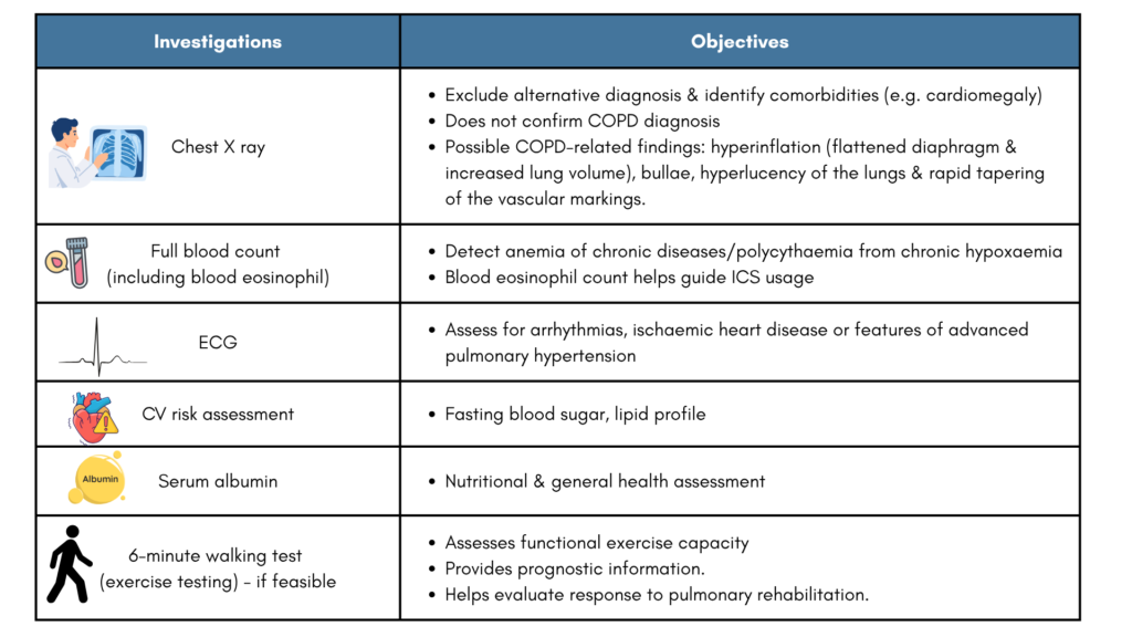
Consider referral for further test
Refer for further assessment when symptoms remain unexplained or disproportionate to airflow obstruction.
Further test to consider,
a) Full lung-function testing, including lung volumes and DLCO
b) Chest CT for suspected alternative disease, lung-volume reduction assessment or eligible lung-cancer screening
c) ABG if :
- FEV1 < 40% predicted
- SpO2 <92% on pulse oximetry
- Clinical signs of resp. failure or cor pulmonale
d) Alpha-1 antitrypsin deficiency (AATD) screening
- The Malaysia CPG recommends testing :
– In young COPD pt (< 45 y/o) or
– Those who have a strong family history ) - GOLD recommends screening for AATD in all patients diagnosed with COPD.
Management – general, non-pharmacological, pharmacological
General – for all COPD patients
1. Risk-factor reduction
Smoking cessation is the most effective and cost-effective intervention for slowing COPD progression. Offer support to every smoker using the 5As approach:
Ask → Advise → Assess → Assist → Arrange follow-up
Treatment options include:
- Behavioural counselling—individual or group
- Nicotine replacement therapy
- Varenicline
- Bupropion
- Nortriptyline
Combining counselling with pharmacotherapy is generally most effective.
Reduce household pollutant exposure through adequate ventilation, cleaner cooking fuels and non-polluting stoves
2. Vaccination
a) Year Influenza vaccination
b) SARS-CoV-2 (COVID-19)
c) Pneumococcal vaccination
- For all COPD patients at least once in their lifetime & have it repeated ≥ 5 years (with a max of 2 doses in one’s lifetime)
- If not vaccinated previously/history unknown, current recommendation:
– PCV 13 or PCV 15 followed by PPSV23 or one-dose PCV 20, or one-dose PCV 21.
– GOLD recommend the use of PCV 20 or 21 in patients with COPD.
d) RSV : for those ≥ 50 years &/or with chronic heart or lung disease
e) Tdap (if not previously vaccinated)
f) Zoster (Shingles) vaccination – for people with COPD aged > 50 years
3. Patient Education & Self-Management
Provide individual or group education covering:
- Smoking cessation & basic understanding of COPD
- Strategies for managing breathlessness
- Exacerbation action plans & when to seek help
- Advance care planning in advanced COPD
Education should extend beyond simply providing information. Support patients in developing practical, sustainable self-management skills.
Differential diagnosis of COPD
- Bronchial asthma
- CCF
- PTB
- Bronchiectasis
- Diffuse parenchymal lung disease
- Pulmonary vascular disease
Pharmacological management of stable COPD (CPG guidelines)
We’ll be discussing the pharmacological management based on 2 guidelines – Malaysia CPG & GOLD 2026 guidelines
The Malaysia CPG was last updated in 2009, so it may be useful to incorporate the latest evidence-based recommendations from GOLD 2026 to keep ourselves up to date.
Bronchodilator remains the mainstay of initial treatment.
Malaysia CPG
The algorithm below seems to be quite compact & “busy” looking. A summarized version (based on my interpretation) are as follows:
- Mild COPD: SABA/SAMA or combination of both may be prescribed.
- Moderate to very severe COPD:
– Either LABA or LAMA, or combination of both.
– If symptoms persist, then consider ICS/LABA+LAMA ± theophylline.
In places where LABA/LAMA may not be available (resource-limited settings), may consider to start SABA/SAAC combination regularly in moderate to severe COPD.
In GOLD 2026 guidelines : Addition of ICS should take into account certain factors, which will be discussed shortly. Also theophylline is no longer recommended due to significant side effects.
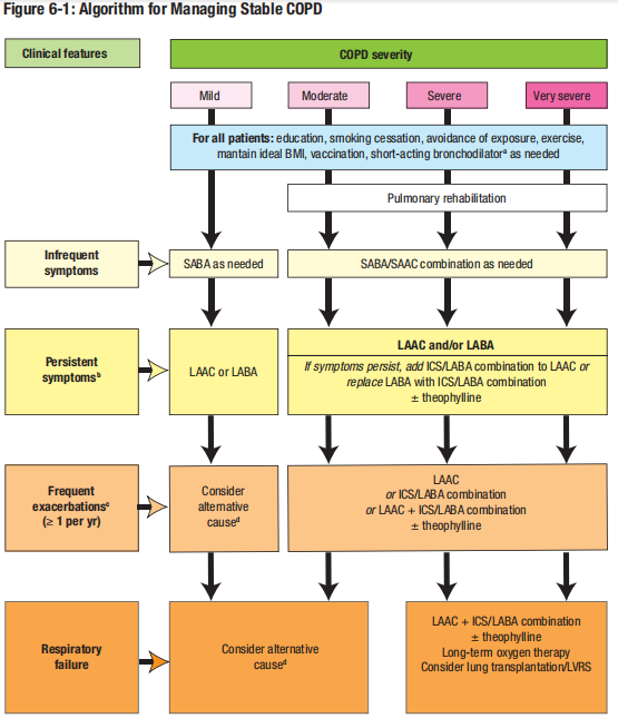
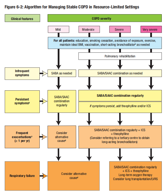
Note :
- ICS dose per day should be at least 500 µg of fluticasone or 800 µg of budesonide
- All COPD patients (regardless of severity) should be prescribed SABA or SABA/SAAC combination (e.g. Berodual, Combivent) for PRN use.
- Persistent symptoms = Need for rescue bronchodilators > 2 times per week
- Frequent exacerbations = ≥ 1 exacerbation requiring systemic corticosteroids +- antibiotics &/or hospitalization over the past 1 year.
- Frequent exacerbations are uncommon in mild COPD, while respiratory failure is uncommon in mild to moderate COPD. If either occurs, investigate for alternative causes, even when COPD is firmly established.
GOLD 2026
Treatment can generally be approached by whether patient is treatment naive or not.
Treatment naive patients
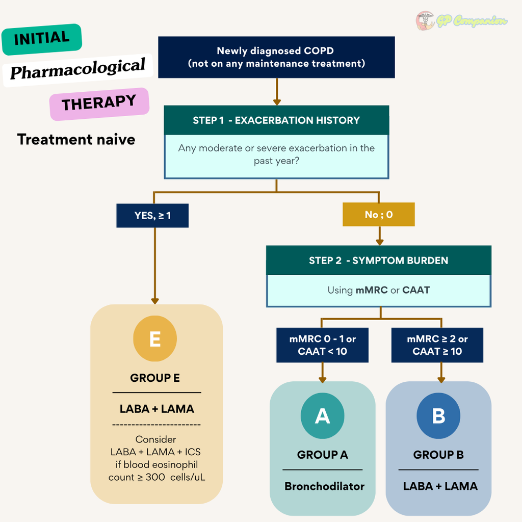
Pharmacological treatment is based on exacerbation history & symptoms burden (mMRC, CAAT) which categorized patients into Group A, B or E as mentioned before.
Group A
- Either short- or a long- acting bronchodilator
- If available & affordable, a long-acting bronchodilator is preferred except in patients with very occasional breathlessness.
Group B
- LABA + LAMA combination (recommended)
- If LABA + LAMA combination is not considered appropriate (cost, availability, side-effects), then either one can be used for symptom relief based on patient’s perception of symptom relief
Group E
- LABA + LAMA combination (preferred)
- Use of LABA + ICS is not encouraged. If ICS is indicated, LABA + LAMA + ICS (triple therapy) is preferred.
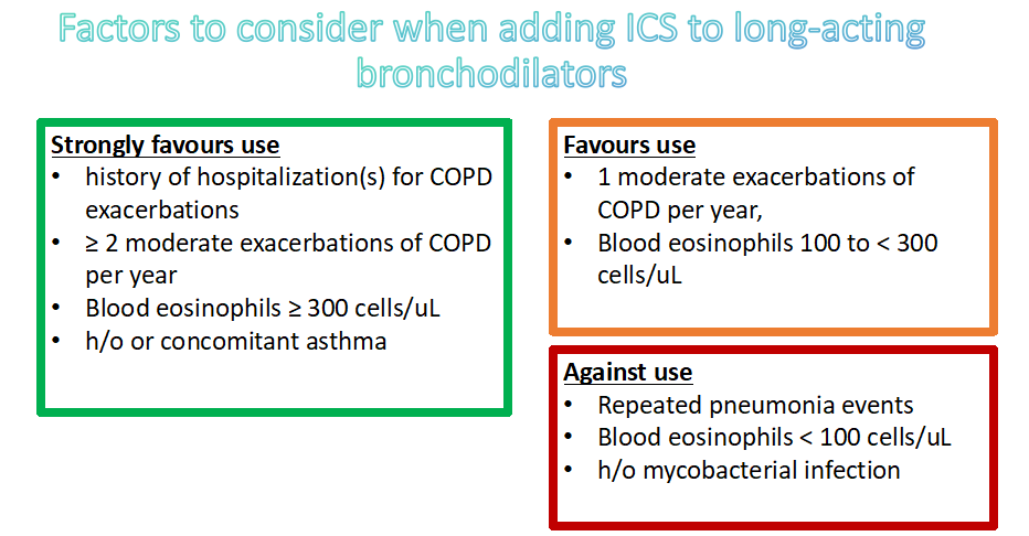
- Consider triple therapy as initial therapy if eosinophil counts ≥ 300 cells/uL
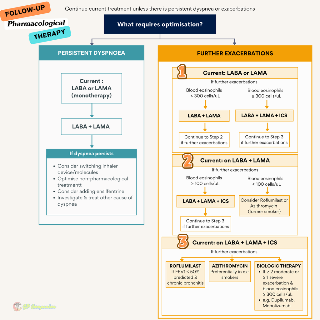
Follow up pharmacotherapy
Based on 2 key treatable traits :
i. Persistence of dyspnea
ii. Occurrence of exacerbations
Consider the predominant treatable trait to target (dyspnea or exacerbations). Use the exacerbation pathway if both exacerbations & dyspnea need to be targeted.
Follow up should be guided by the principles of review, assess & adjust if necessary.
- Review : symptoms (dyspnea), exacerbation risk (previous history), blood eosinophils
- Assess : adherence, inhaler technique & role of non-pharmacological treatment
- Adjust : pharmacological treatment (escalate or de-escalate), switching inhaler device if indicated.
Consideration for adding ICS
Regarding LABA + ICS
Use of LABA + ICS routinely is not encouraged.
If there is an indication for ICS then LABA + LAMA + ICS has been shown to be superior to LABA + ICS and is therefore the preferred choice.
In patients with COPD who have no clinical features of asthma, if they were on ICS + LABA (for whatever reason), continuation of LABA/ICS therapy may be considered when symptoms and exacerbations remain well controlled.
However, if the patient has :
- Further exacerbations – escalate to triple therapy if the blood eosinophil count is ≥ 100 cells/uL or switched to LABA + LAMA if it is ≤ 100 cells/uL.
- Major symptoms – change to LABA + LAMA or triple therapy depending on previous treatment response to ICS.
Non-pharmacological management
1. Pulmonary Rehabilitation
A personalised programme combining exercise, education and self-management support to improve physical function and psychological well-being.
Consider for: Patients with significant symptoms or exacerbation risk, particularly Groups B and E.
Assessment includes:
- Medical history, examination and post-bronchodilator spirometry
- Exercise capacity, such as the 6-minute walk or shuttle walk test
- Breathlessness, fatigue and health status
- Respiratory and lower-limb muscle strength when muscle wasting is present
- Patient goals, daily limitations, expectations and understanding of COPD
2. Long-Term Oxygen Therapy (> 15 hours daily)
Improves survival in severe chronic resting hypoxaemia but not moderate resting or exercise-induced desaturation.
Indications:
- PaO₂ ≤ 7.3 kPa (55 mmHg) or SaO₂ ≤88% ± hypercapnea (confirmed twice over a 3 week period); or
- PaO₂ 7.3 – 8.0 kPa (55–60 mmHg) or SaO₂ 89% with pulmonary hypertension, peripheral oedema or haematocrit >55%
Contraindications
- PaO₂ >60 mmHg without chronic hypoxia
- Active smoking
- COPD treatment is not yet optimized (e.g. bronchodilators, treatment for any respiratory infection, etc)
- Poor adherence is anticipated
Assessment should not be done during exacerbation/recovery period of exacerbation. ABG should be made on 2 occasions when the pt is in a stable condition & on optimal treatment.
After starting LTOT, reassess clinically and with ABG after 2–3 months, then at least annually or accordingly.
3. Ventilatory Support
Consider long-term NIV in selected patients with marked daytime hypercapnia, particularly after recent hospitalization.
Use CPAP when COPD coexists with obstructive sleep apnoea.
4. Nutrition
Goal: To maintain healthy weight & BMI (22 – 27 kg/m2) + keep serum albumin > 35 g/L; Prevent both cachexia & obesity.
5. Surgical intervention – Lung volume reduction (e.g. bullectomy etc.), Lung transplantation in indicated patients
Follow up visit – Stable COPD
- Exposure to risk factors, esp. tobacco smoke & their willingness to stop.
- Any new/worsening symptoms.
- Any complications e.g. respiratory failure/cor pulmonale
- Body weight & BMI – provide information on the nutritional status of the pt.
- Spirometry measurement if symptoms worsen.
- Pharmacotherapy & other medical treatment: effectiveness, side effects, compliance, inhaler techniques.
- Exacerbation history
- Any hospitalization, including duration of stay & any use of invasive/non-invasive ventilation
- Patient education
When to refer for a stable COPD patient?
- When the diagnosis is in doubt.
- For spirometry testing when such a facility is not available on site.
- Onset of cor pulmonale.
- Assessment & prescribing of LTOT.
- When there is a rapid decline of FEV1 indicating severity of the disease.
- Pt aged < 40 y/o in whom an underlying genetic predisposition e.g. alpha-1 antitrypsin deficiency is suspected.
- When access to certain drugs/medication is a problem.