
Chronic Kidney Disease
Definition
eGFR < 60 mL/min/1.73 m2 that is present for more than 3 months +/- evidence of kidney damage
OR
Evidence of kidney damage for more than 3 months +/- eGFR < 60 mL/min/1.73 m2
Markers of Kidney Damage
- Albuminuria
- Urine sediment abnormalities
- Electrolyte and other abnormalities due to tubular disorders
- Abnormalities detected by histology
- Structural abnormalities detected by imaging
- History of kidney transplantation
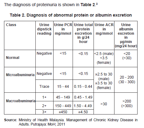
Diagnosis of proteinuria
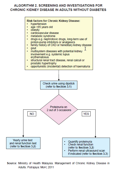
Who to screen for CKD?
- Age > 65 years old
- Obesity CV disease
- Metabolic syndrome (pt with DM &/or HTN should be screened at least yearly)
- Drugs e.g. nephrotoxic drugs, long-term use of PPI or analgesics (e.g. NSAIDs, COX-2 inhibitors)
- Family h/o CKD or hereditary disease
- Gout
- Multisystem diseases with potential kidney involvement e.g. SLE
- Structural renal disease, renal calculi or prostatic hypertrophy
- Incidental detection of hematuria or proteinuria
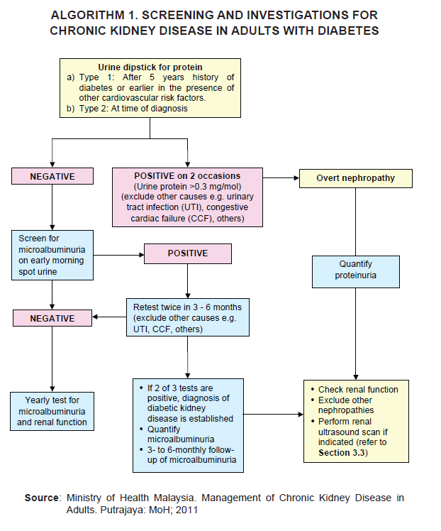
Screening algorithm
Approach to haematuria
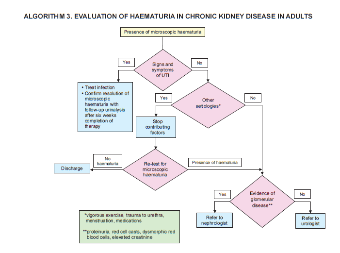
- A positive dipstick test for blood on 2 out of 3 occasions warrant a full microscopic examination.
- Urine microscopy can be used to differentiate hematuria of glomerular or non-glomerular origin.
- Some common causes of persistent microscopic hematuria: infection, glomerulonephritis, renal calculi, malignancy & other forms of kidney damage